Beranda
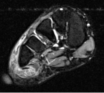
/ Foot Muscles Mri : Ankle and Foot | Radiology Key : Accessory soleus, peroneus quartus and the flexor digitorum longus accessorius.
Foot Muscles Mri : Ankle and Foot | Radiology Key : Accessory soleus, peroneus quartus and the flexor digitorum longus accessorius.
Insurance Gas/Electricity Loans Mortgage Attorney Lawyer Donate Conference Call Degree Credit Treatment Software Classes Recovery Trading Rehab Hosting Transfer Cord Blood Claim compensation mesothelioma mesothelioma attorney Houston car accident lawyer moreno valley can you sue a doctor for wrong diagnosis doctorate in security top online doctoral programs in business educational leadership doctoral programs online car accident doctor atlanta car accident doctor atlanta accident attorney rancho Cucamonga truck accident attorney san Antonio ONLINE BUSINESS DEGREE PROGRAMS ACCREDITED online accredited psychology degree masters degree in human resources online public administration masters degree online bitcoin merchant account bitcoin merchant services compare car insurance auto insurance troy mi seo explanation digital marketing degree floridaseo company fitness showrooms stamfordct how to work more efficiently seowordpress tips meaning of seo what is an seo what does an seo do what seo stands for best seotips google seo advice seo steps, The secure cloud-based platform for smart service delivery. Safelink is used by legal, professional and financial services to protect sensitive information, accelerate business processes and increase productivity. Use Safelink to collaborate securely with clients, colleagues and external parties. Safelink has a menu of workspace types with advanced features for dispute resolution, running deals and customised client portal creation. All data is encrypted (at rest and in transit and you retain your own encryption keys. Our titan security framework ensures your data is secure and you even have the option to choose your own data location from Channel Islands, London (UK), Dublin (EU), Australia.
Foot Muscles Mri : Ankle and Foot | Radiology Key : Accessory soleus, peroneus quartus and the flexor digitorum longus accessorius.. Check the tendons using the four quadrant approach; With a muscle injury, for example, mri images often show a bright signal indicating that there is more water in the muscle, which is a sign of injury. An ankle mri also offers a look at the bones of the lower leg that help make up the ankle joint, such as the tibia and fibula, as well as the muscles of the foot. This test uses radio waves and a strong magnetic field to create detailed images. There are 10 intrinsic muscles located in the sole of the foot.
Screen for effusion and look at the joint capsule for thickening. A case report and review of anatomy. Magnetic resonance imaging of anomalous leg muscles: An ankle mri also offers a look at the bones of the lower leg that help make up the ankle joint, such as the tibia and fibula, as well as the muscles of the foot. The machine uses radio waves and a magnetic field to generate images of the inside of the extremity in order to diagnose problems with the muscles, bones, joints, nerves, or blood vessels.
Baxter's Nerve (First Branch of the Lateral Plantar Nerve ... from radsource.us Posted by radiologyer at 8:12 am. The studies were performed on a variety of magnets ranging from 0.2 to 1.5 t between march 15 and july 22, 2006. Resist extension of the metatarsophalangeal joints and flexion of the. Lateral and medial processes of calcaneal tuberosity, and band of connective tissue connecting calcaneus with base of metatarsal v; Your doctor, with the help of a radiologist, can then examine these images to determine whether there is anything wrong with your foot or. An ankle mri also offers a look at the bones of the lower leg that help make up the ankle joint, such as the tibia and fibula, as well as the muscles of the foot. Due to complexity of the plantar intrinsic foot muscles, little is known about their muscle architecture in vivo. 9 yao l, do hm, cracchiolo a, et al.
Screen for effusion and look at the joint capsule for thickening.
Flexors on the medial side. The machine uses radio waves and a magnetic field to generate images of the inside of the extremity in order to diagnose problems with the muscles, bones, joints, nerves, or blood vessels. Plantar plate of the foot: 9 yao l, do hm, cracchiolo a, et al. Chronic plantar fasciitis may be accompanied by muscle atrophy of plantar intrinsic foot muscles and tibialis posterior compromising the dynamic support of the foot prolonging the injury. Fractures or breaks in the lower portion of the tibia and fibula will show up. Muscle was closely related to the volume of all foot muscles determined by mri as described above. Screen for effusion and look at the joint capsule for thickening. Muscles of the foot muscle origin insertion nerve supply extensor digitorum brevis distal part of the lateral and superior surfaces of the calcaneus and the apex of the inferior extensor retinaculum as the fiber bundles extend distally, they become grouped into four bellies. The insufficiency of the ligaments and muscles of the foot sole often lead to foot deformities. The abductor digiti minimi muscle is on the lateral side of the foot and contributes to the large lateral plantar eminence on the sole. Proximal margin and deep surface of. They act collectively to stabilise the arches of the foot, and individually to control movement of the digits.
There are 10 intrinsic muscles located in the sole of the foot. Due to complexity of the plantar intrinsic foot muscles, little is known about their muscle architecture in vivo. Those fibers of the most medial and largest belly are… They are named extensor digitorum brevis and extensor hallucis brevis. 23 it can originate as a separate muscle from the fibula or from the peroneus brevis or longus muscles and inserts onto the peroneal tubercle or retrotrochlear eminence of the calcaneus.
MRI anatomy of hip joint | free MRI axial hip anatomy from mrimaster.com The three plantar interossei muscles adduct the 3 rd, 4 th and 5 th toes toward the long axis through the 2 nd toe. All the muscles are innervated either by the medial plantar nerve or the lateral plantar nerve, which are both branches of the tibial nerve. 9 yao l, do hm, cracchiolo a, et al. These include plantar fibromatosis, haemangioma, lipoma, pvns/gct tendon sheath and synovial chondromatosis. Muscle anatomy trivia 12 photos of the muscle anatomy trivia muscle anatomy trivia, human muscles, muscle anatomy trivia The muscles lie within a flat fascia on the dorsum of the foot (fascia dorsalis pedis) and are innervated by the deep fibular or peroneal nerve. Related posts of foot muscle anatomy mri muscle anatomy trivia. Check the syndesmosis, the lateral and medial ligaments.
Check the syndesmosis, the lateral and medial ligaments.
Posted by radiologyer at 8:12 am. Upper two thirds of the medial margin and proximal margin of the patella, medial condyle of the tibia, and investing deep fascia of the leg with the tendons of vastus intermedius, lateralis, and rectus, and through the patellar ligament onto the front of the tibial tuberosity. Extensor hoods and bases of proximal phalanges of toes iii to v action: A case report and review of anatomy. They are named extensor digitorum brevis and extensor hallucis brevis. This imaging technique assesses the ligaments and tendons, neurovascular structures ( tarsal tunnel and plantar fascia), and the osseous structures (19). 23,25 mri at the level of the malleolus demonstrates the muscle as. Anatomy of the whole human body : The three plantar interossei muscles adduct the 3 rd, 4 th and 5 th toes toward the long axis through the 2 nd toe. The machine uses radio waves and a magnetic field to generate images of the inside of the extremity in order to diagnose problems with the muscles, bones, joints, nerves, or blood vessels. Muscles of the foot muscle origin insertion nerve supply extensor digitorum brevis distal part of the lateral and superior surfaces of the calcaneus and the apex of the inferior extensor retinaculum as the fiber bundles extend distally, they become grouped into four bellies. Lateral side of base of proximal phalanx of little toe. Related posts of foot muscle anatomy mri muscle anatomy trivia.
The insufficiency of the ligaments and muscles of the foot sole often lead to foot deformities. A case report and review of anatomy. Shoulder elbow wrist finger thumb. The three plantar interossei muscles adduct the 3 rd, 4 th and 5 th toes toward the long axis through the 2 nd toe. Magnetic resonance imaging, otherwise known as mri, uses a combination of magnetic fields and radio waves to take images of the internal structures of your body.
MRI proves tennis leg injuries are more than just a racket from www.auntminnie.com Mri findings of acute turf toe: • muscle edema is seen secondary to multiple etiologies including trauma, infectious and inflammatory processes, autoimmune disorders, neoplasms, and denervation injuries • on mri muscle edema is characterized by increase in free water within the muscle • muscle edema is seen on mri as increased signal on fluid sensitive sequences t2 fs Certain soft tissue tumours are identifiably benign because of their signal characteristics, morphology and/or location. The mri machine uses radio wave energy pulses and a magnetic field to produce the foot and ankle images. These include plantar fibromatosis, haemangioma, lipoma, pvns/gct tendon sheath and synovial chondromatosis. A case report and review of anatomy. In addition, any strains or tears of the foot muscles will also be visible. Like the fingers, the toes have flexor and extensor muscles that power their movement and play a large.
This test uses radio waves and a strong magnetic field to create detailed images.
Mri is an ideal method for identifying areas of muscle atrophy and fatty infiltration. The muscles lie within a flat fascia on the dorsum of the foot (fascia dorsalis pedis) and are innervated by the deep fibular or peroneal nerve. Accessory muscles are isointense to skeletal muscle on all pulse sequences, and can insert by fleshy muscular or tendinous insertions. Foot and ankle mri — what you should know. Like the fingers, the toes have flexor and extensor muscles that power their movement and play a large. Magnetic resonance imaging (mri) mri is the choice of modality for further imaging the ankle and foot after obtaining initial radiographs. With a muscle injury, for example, mri images often show a bright signal indicating that there is more water in the muscle, which is a sign of injury. Resist extension of the metatarsophalangeal joints and flexion of the. In addition, an image of all the muscles of the back and plantar part of the foot, all tendons and tendon ligaments, blood vessels and nerves are obtained. Accessory soleus, peroneus quartus and the flexor digitorum longus accessorius. Electromyography (emg) and nerve conduction studies measure electrical activity in the muscles and nerves. Muscle anatomy trivia 12 photos of the muscle anatomy trivia muscle anatomy trivia, human muscles, muscle anatomy trivia Fractures or breaks in the lower portion of the tibia and fibula will show up.